Skip to content.

The Connection Between Diabetes, Arteries, and Limb Health

A small cut on your foot, aching legs after a short walk, or a sore that takes longer than expected to heal might not seem like reasons for concern. However, for people living with diabetes, these changes can sometimes point to something more serious happening beneath the surface. 

Diabetes can affect how well blood circulates throughout the body, increasing the risk of narrowed arteries, poor circulation, and peripheral artery disease (PAD). When blood flow to the legs and feet becomes restricted, even minor injuries can develop into difficult-to-heal wounds and potentially serious limb complications. 

Understanding how diabetes affects artery health is essential for recognizing early warning signs, protecting circulation, and reducing the risk of long-term damage. In this article, we’ll explore the connection between diabetes and peripheral artery disease, why lower limb health deserves particular attention, and what you can do to support healthy circulation.

How Does Diabetes Affect Your Arteries?

Diabetes occurs when the body has difficulty producing or using insulin, causing blood glucose levels to remain higher than normal. When elevated blood sugar persists over time, it can damage the lining of blood vessels and contribute to changes that affect circulation.

This damage can make it easier for fatty deposits, cholesterol, and other substances to build up inside the arteries. As these deposits accumulate, the arteries can become narrower and less flexible, restricting the amount of blood that reaches different parts of the body.

Several factors can further increase the risk of vascular complications in people with diabetes, including:

  • High blood pressure: Places additional strain on artery walls and contributes to vascular damage.
  • High cholesterol: Can accelerate plaque buildup inside the arteries.
  • Smoking: Damages blood vessels and further restricts circulation.
  • Long-term elevated blood sugar: Can contribute to ongoing damage to blood vessels and nerves.

Together, these factors can significantly increase the risk of developing circulation problems, particularly in the lower extremities.

The Link Between Diabetes and Peripheral Artery Disease (PAD)

Peripheral artery disease is a common circulatory condition that affects arteries outside the heart and brain, most often those supplying blood to the legs. It typically develops when plaque accumulates inside the arteries, restricting blood flow.

People with diabetes have a higher risk of developing PAD, partly because prolonged high blood sugar can damage blood vessels and contribute to atherosclerosis, the buildup of plaque inside artery walls.

When circulation to the legs and feet becomes restricted, the body may struggle to deliver enough oxygen-rich blood to support everyday activities and maintain healthy tissue.

Common signs of PAD include:

  • Leg pain when walking: Cramping, aching or fatigue in the calves, thighs, or hips during physical activity, often improving with rest.
  • Changes in skin temperature or color: One foot or leg may feel cooler than the other or appear unusually pale or discolored.
  • Slow-healing wounds: Cuts, sores, or ulcers on the feet and legs may take longer to heal.
  • Reduced hair growth: Less hair may develop on the lower legs and feet.
  • Weak or absent pulses: Blood flow to the feet may be reduced.

Not everyone with PAD experiences noticeable symptoms. This is particularly important for people with diabetes, who may also have nerve damage that makes it harder to recognize pain or injuries.

Why Foot Health Is Especially Important When You Have Diabetes

The combination of reduced circulation and diabetic neuropathy (nerve damage) can make even minor foot injuries more concerning.

When blood flow is limited, the body has a harder time delivering the oxygen and nutrients needed for tissue repair. At the same time, nerve damage can reduce sensation, meaning a person may not notice a blister, cut, or developing sore until it becomes more serious.

Without proper attention, these seemingly minor problems can lead to:

  • Foot ulcers: Open sores that develop when damaged skin struggles to heal.
  • Infections: Bacteria can enter through breaks in the skin, potentially leading to more serious complications.
  • Tissue damage: Severely reduced blood flow can cause tissue to deteriorate, sometimes resulting in gangrene.
  • Limb-threatening complications: In advanced cases, untreated circulation problems and infections can increase the risk of amputation.

This is why monitoring both nerve health and circulation is so important for people with diabetes. A wound that appears small should never be dismissed simply because it doesn’t hurt.

How to Protect Your Artery and Limb Health

Managing diabetes involves more than keeping blood sugar within your target range. Taking steps to support circulation and identify potential vascular problems early can help reduce the risk of complications.

Here are several habits worth incorporating into your routine:

  1. Check Your Feet Every Day

Look for cuts, blisters, swelling, redness, discoloration, temperature changes, or sores that aren’t healing. Pay attention to the areas between your toes and the soles of your feet, too.

  1. Keep Blood Sugar Under Control

Work with your healthcare provider to manage blood glucose levels. Consistent diabetes management can help reduce the risk of blood vessel and nerve damage.

  1. Stay Physically Active

Regular movement, including walking and other activities approved by your healthcare provider, can support circulation and overall cardiovascular health.

  1. Avoid Smoking

Smoking damages blood vessels, contributes to plaque buildup and further restricts blood flow. Quitting can be an important step toward protecting your arteries.

  1. Manage Other Cardiovascular Risk Factors

Keep track of your blood pressure and cholesterol, follow your prescribed treatment plan and attend routine medical appointments.

  1. Don’t Ignore Changes in Your Legs or Feet

New or recurring leg pain, numbness, changes in skin color, or wounds that aren’t healing deserve medical attention. Don’t assume these changes are simply part of aging or diabetes.

If you experience sudden severe leg or foot pain, a suddenly cold or pale limb, or sudden loss of sensation or movement, seek emergency medical care.

When Should You See a Vascular Specialist?

If you have diabetes, you may benefit from discussing your vascular health with a specialist, particularly if you have additional risk factors for peripheral artery disease or notice changes in your legs or feet.

A vascular specialist can evaluate circulation, identify potential arterial blockages and recommend appropriate next steps. Depending on your symptoms and medical history, an evaluation may include:

  • Ankle-brachial index (ABI): Compares blood pressure in the ankle and arm to help identify reduced blood flow. Diabetes-related artery calcification can sometimes affect results, so additional testing may be needed.
  • Vascular ultrasound: Uses sound waves to assess blood flow and identify areas of narrowing or blockage.
  • Additional imaging: Depending on the findings, further imaging may help determine the location and severity of arterial disease.

Treatment depends on the extent of the condition and individual health needs. Options may include medication, lifestyle modifications, or procedures designed to improve blood flow, such as angioplasty or other revascularization techniques.

Early evaluation can help identify circulation problems before they progress to more serious complications.

Protect Your Vascular Health with The Vascular Care Group

Living with diabetes means paying attention to more than your blood sugar. Your arteries, circulation, and limb health all play an important role in maintaining your mobility and quality of life. Recognizing potential warning signs and addressing circulation concerns early can help you take a more proactive approach to your health.

At The Vascular Care Group, our experienced vascular specialists provide comprehensive care for patients with peripheral artery disease and other vascular conditions. With convenient locations throughout Connecticut, Massachusetts and Maine, we help patients understand their vascular health and explore appropriate treatment options.

If you’re experiencing leg pain, changes in circulation, or wounds that aren’t healing, don’t wait for symptoms to worsen. Contact The Vascular Care Group to schedule an evaluation and take the next step toward healthier circulation.


 

What Does Your Carotid Artery Have to Do With Stroke Risk?

You can take steps to protect your heart health without realizing that another set of blood vessels deserves your attention: your carotid arteries. Located on both sides of your neck, these major arteries carry oxygen-rich blood to your brain, making their health essential for normal brain function. 

When plaque builds up inside the carotid arteries, the resulting narrowing, known as carotid artery disease, can restrict blood flow and increase the risk of a stroke. In some cases, pieces of plaque or blood clots can travel to the brain and block circulation, potentially causing lasting neurological damage. What makes carotid artery disease particularly concerning is that it develops without obvious symptoms, leaving many people unaware of their risk. 

So, how does your carotid artery affect your chances of having a stroke, and what can you do to protect yourself? Understanding the relationship between carotid artery disease and stroke risk, including common risk factors, potential warning signs, and available treatment options, can help you make informed decisions about your vascular health. 

At The Vascular Care Group, patients can access specialized vascular care at locations throughout Connecticut, Massachusetts, and Maine, with evaluations tailored to their individual needs.

What Are the Carotid Arteries, and Why Do They Matter?

Your carotid arteries are two major blood vessels located on either side of your neck. They carry oxygen-rich blood from your heart to your brain, supporting everything from movement and memory to speech and concentration. When these arteries become narrowed or blocked, your brain may not receive the blood supply it needs.

Carotid artery disease most often develops through atherosclerosis, a condition in which plaque made of cholesterol, fat, calcium, and other substances accumulates along artery walls. Over time, this buildup can narrow the passageway for blood or contribute to blood clot formation.

The connection between carotid artery disease and stroke risk comes down to two primary concerns:

  • Reduced blood flow: Significant narrowing can limit the amount of blood reaching the brain.
  • Plaque or clot blockage: Pieces of plaque or blood clots can break loose, travel to the brain, and block a smaller artery, causing an ischemic stroke.

Understanding how carotid artery disease develops is an important first step toward recognizing your risk and discussing appropriate prevention with your healthcare provider.

What Increases Your Risk of Carotid Artery Disease?

Carotid artery disease often develops gradually, influenced by health conditions, lifestyle habits, and factors you cannot control. Some of the same factors that contribute to plaque buildup also increase your overall risk of stroke.

Common risk factors include:

  • High cholesterol: Elevated cholesterol can contribute to plaque accumulation inside the arteries.
  • High blood pressure: Persistent pressure on artery walls can damage blood vessels and increase cardiovascular risk.
  • Smoking: Tobacco use damages blood vessels and promotes the development of atherosclerosis.
  • Diabetes: Elevated blood sugar can damage blood vessels and increase the likelihood of vascular complications.
  • Age and family history: The risk of atherosclerosis generally increases with age, and a family history of cardiovascular disease may contribute to your risk.
  • Physical inactivity and an unhealthy diet: These habits can contribute to high cholesterol, high blood pressure, and other conditions associated with arterial disease.

Having one or more risk factors does not necessarily mean you have carotid artery disease. However, discussing your cardiovascular health with a healthcare provider can help determine whether further evaluation is appropriate.

Can Carotid Artery Disease Develop Without Symptoms?

One of the biggest challenges with carotid artery disease is that it may not cause noticeable symptoms in its early stages. Some people discover they have narrowed carotid arteries during an evaluation for another medical concern, while others are diagnosed after experiencing a transient ischemic attack (TIA) or stroke.

A TIA, sometimes called a mini-stroke, occurs when blood flow to part of the brain is temporarily disrupted. Symptoms may resolve quickly, but a TIA is still a medical emergency and a warning sign that requires immediate evaluation.

Watch for sudden symptoms such as:

  • Weakness or numbness in the face, arm, or leg, especially on one side of the body
  • Difficulty speaking or understanding speech
  • Sudden vision changes in one or both eyes
  • Dizziness, loss of balance, or difficulty walking
  • A sudden, severe headache with no known cause

If you or someone nearby experiences possible stroke symptoms, call 911 immediately, even if the symptoms disappear. Do not wait for a medical appointment or attempt to drive yourself to the hospital.

How Is Carotid Artery Disease Diagnosed?

If your medical history, symptoms, or examination suggest carotid artery disease, your healthcare provider may recommend additional testing. A diagnosis helps determine how significantly the artery is narrowed and whether treatment is needed.

Common evaluation methods include:

  • Physical examination: A provider may listen to the carotid arteries for an unusual whooshing sound, called a bruit, which can indicate turbulent blood flow. However, its absence does not rule out disease.
  • Carotid duplex ultrasound: This noninvasive test uses sound waves to evaluate blood flow and identify narrowing in the carotid arteries.
  • Additional imaging: Depending on the findings, CT angiography or MR angiography may provide more detailed images of the blood vessels.

Not everyone needs carotid artery screening. Your provider can assess your individual risk and determine whether testing is appropriate.

What Treatments Can Help Lower Stroke Risk?

Treatment for carotid artery disease depends on the degree of narrowing, whether you have experienced symptoms, and your overall health. The goal is to reduce the risk of stroke while addressing the underlying vascular disease.

Depending on your circumstances, treatment may include:

  • Medication: Medicines to manage cholesterol, blood pressure, or clotting risk may be recommended.
  • Lifestyle changes: Quitting smoking, staying physically active, eating a balanced diet, and managing chronic conditions can support vascular health.
  • Carotid endarterectomy: This surgical procedure removes plaque from the affected artery.
  • Carotid angioplasty and stenting: In selected cases, a catheter-based procedure is used to widen the narrowed artery and place a stent to help keep it open.

Procedures are not necessary for everyone with carotid artery disease. A vascular specialist can explain the potential benefits and risks of available options and recommend an approach based on your individual needs.

Protect Your Vascular Health With The Vascular Care Group

Understanding the relationship between carotid artery disease and stroke risk can help you take a more proactive approach to your health. If you have concerns about your risk factors, have been diagnosed with narrowed carotid arteries, or need follow-up care after a vascular evaluation, speaking with a specialist can help you understand your options.

The Vascular Care Group provides specialized vascular care at locations throughout Connecticut, Massachusetts, and Maine. Our team evaluates and treats a range of vascular conditions, helping patients understand their diagnoses and make informed decisions about their care.

Visit our locations page to find a convenient office and learn more about the vascular services available near you.


What Happens After a DVT? Understanding Long-Term Vein Health

A deep vein thrombosis (DVT) can be frightening, and having the blood clot treated is an important step toward protecting your health. But what happens after a DVT has been resolved? For some people, the effects can linger long after the initial clot is gone, leading to persistent leg swelling, discomfort, skin changes, or an increased risk of developing another blood clot. These symptoms may signal ongoing issues with vein function and circulation that deserve medical attention. 

Understanding how a DVT can affect your long-term vein health, recognizing potential complications, and knowing when to seek follow-up care can help you take informed steps toward protecting your vascular health. 

Whether you have recently been diagnosed with deep vein thrombosis or are navigating life after treatment, knowing what to expect can make a meaningful difference in your recovery and long-term well-being.

How Does a DVT Affect Your Veins?

Deep vein thrombosis occurs when a blood clot forms in a deep vein, most often in the leg. These veins carry blood back toward the heart, working against gravity with help from small valves that keep blood flowing in the right direction.

A DVT can interfere with this process in two ways:

  • Restricted blood flow: A clot can partially or completely block a vein, making it harder for blood to circulate normally.
  • Damage to vein valves: Even after a clot is treated, inflammation and damage to the vein walls or valves can interfere with circulation.

Although treatment helps reduce the risk of serious complications, the affected vein may not return to its previous condition. This is why follow-up care matters, even after the initial symptoms improve.

What Is Post-Thrombotic Syndrome?

One of the most common long-term complications of DVT is post-thrombotic syndrome (PTS). This condition develops when a previous blood clot damages the veins or their valves, allowing blood to pool in the affected leg instead of flowing efficiently back toward the heart.

Symptoms can develop weeks or months after a DVT and may include:

  • Persistent swelling in the lower leg or ankle
  • Aching, cramping, or heaviness in the affected leg
  • Pain that worsens after standing or walking for extended periods
  • Skin discoloration, itching, or thickening around the ankle
  • Venous ulcers in more severe cases

Symptoms range from mild discomfort to more significant changes that interfere with everyday activities. If you notice ongoing leg swelling or discomfort after a DVT, a vascular specialist can evaluate your circulation and recommend appropriate care.

Can You Develop Another Blood Clot After a DVT?

Yes. Having a DVT increases your risk of developing another blood clot, particularly if certain risk factors remain present. Although treatment helps prevent complications, it does not eliminate the possibility of recurrence.

Factors that may increase your risk include:

  • Prolonged sitting or immobility, including during long trips
  • Recent surgery or hospitalization
  • Certain cancers or medical conditions
  • A personal or family history of blood clots
  • Some hormone-based medications or treatments

Recognizing your individual risk factors can help you and your healthcare provider determine the best approach to prevention. Depending on your circumstances, this may involve medication, regular movement, compression garments when recommended, or additional monitoring.

Seek emergency medical attention if you develop sudden shortness of breath, chest pain, coughing up blood, or fainting, as these may indicate a pulmonary embolism, a potentially life-threatening complication in which a clot travels to the lungs. New or worsening leg swelling, warmth, redness, or pain also warrants prompt medical evaluation.

How Can You Protect Your Long-Term Vein Health?

Life after a DVT often involves a combination of healthy habits, appropriate medical treatment, and follow-up appointments. While you cannot eliminate every risk factor, there are practical steps that may support circulation and help reduce your risk of future complications.

Consider these strategies:

  • Stay physically active. Walking and other activities recommended by your healthcare provider encourage circulation and help prevent prolonged immobility.
  • Take medications as prescribed. If you have been prescribed anticoagulants, follow your treatment plan and consult your provider before making changes.
  • Break up long periods of sitting. Move your legs regularly and take walking breaks during extended travel or desk work.
  • Discuss compression therapy. Compression stockings may help manage swelling and discomfort for some people, but they should be selected with guidance from your healthcare provider.
  • Keep follow-up appointments. Monitoring your symptoms and vein health can help identify complications that may need further evaluation.

If leg discomfort, swelling, or skin changes persist, do not assume they are simply part of recovery. A vascular evaluation can help determine whether ongoing vein problems are contributing to your symptoms.

When Should You See a Vascular Specialist After a DVT?

Follow-up care is important if you have experienced DVT, especially if symptoms persist or return after treatment. A vascular specialist can review your medical history, assess your symptoms, and determine whether additional testing, such as a vascular ultrasound, may be appropriate.

People with ongoing symptoms may benefit from an individualized treatment plan focused on managing chronic venous problems, improving comfort, and protecting long-term vascular health. The right approach depends on the location and severity of the original clot, any lasting vein damage, and your overall health.

Get Support for Your Long-Term Vein Health

Recovering from a DVT does not always mean your vein health concerns are behind you. Recognizing lingering symptoms, understanding your risk of another blood clot, and seeking appropriate follow-up care can help you make informed decisions about your circulation.

At The Vascular Care Group, our vascular specialists provide evaluation and treatment for venous disease at convenient locations throughout Connecticut, Massachusetts, and Maine. If you have ongoing concerns following a DVT or want to learn more about your vein health, our team can help you explore your options.


PAD Treatment Options: How Doctors Decide Between Traditional Bypass Surgery and Minimally Invasive Procedures

When leg pain, cramping or fatigue start making everyday activities more difficult, it can be hard to know what the next step should be. For people living with peripheral artery disease (PAD), reduced blood flow caused by narrowed or blocked arteries can lead to symptoms that affect everything from walking and exercising to completing routine tasks. The good news is that PAD treatment has advanced significantly, and patients today may have several options depending on their symptoms and the location and severity of their arterial disease. 

From minimally invasive procedures and angioplasty with stent placement to more complex vascular surgery such as bypass, treatment is tailored to the individual rather than following a one-size-fits-all approach. Understanding how vascular specialists evaluate PAD and determine the most appropriate treatment can help patients feel more informed about their options and the path toward improved circulation, mobility and vascular health.

Why PAD Treatment Is Not One-Size-Fits-All

Peripheral artery disease occurs when narrowed or blocked arteries reduce blood flow, most often to the legs and feet. While some people experience mild symptoms, others may develop pain while walking, pain at rest or wounds that are slow to heal.

Before recommending a procedure, a vascular specialist evaluates the full picture. Treatment decisions are based on several factors, including:

  • The severity of symptoms
  • The location and length of the blockage
  • How severely blood flow is restricted
  • Whether PAD affects one artery or multiple arteries
  • The presence of nonhealing wounds or tissue damage
  • A patient’s overall health and medical history
  • Previous PAD treatments or procedures

Diagnostic testing may include an ankle-brachial index, ultrasound or other imaging studies that help the doctor understand where blood flow is being affected. This information helps determine whether conservative treatment, a minimally invasive procedure or surgery may be the most appropriate next step.

When Minimally Invasive PAD Procedures May Be Recommended

For many patients, minimally invasive procedures can restore blood flow without requiring traditional open surgery. These procedures are typically performed through small access points in the blood vessels and may involve specialized catheters, balloons or other devices.

Depending on the patient’s condition, options may include:

  • Angioplasty, which uses a small balloon to widen a narrowed artery
  • Atherectomy, which may be used to remove or modify plaque within an artery
  • Drug-coated balloons, which may help keep certain treated arteries open
  • Other endovascular techniques designed to improve blood flow

Minimally invasive PAD treatment may be considered when the anatomy of the blockage and the patient’s overall condition make an endovascular approach appropriate. These procedures can offer a less invasive alternative to open surgery for certain patients, but the right option depends on the individual.

How Doctors Decide Whether a Stent Is Needed

A stent is a small device placed inside an artery to help keep it open after treatment. Stenting may be recommended when angioplasty alone does not provide enough support or when a treated artery is at risk of narrowing again.

Doctors consider several factors before placing a stent, including:

  • The location of the narrowed or blocked artery
  • The length and characteristics of the blockage
  • How the artery responds to angioplasty
  • Whether the area is likely to remain open without additional support
  • The patient’s symptoms and treatment goals

Stents can be an important part of PAD treatment, but they are not automatically the best choice for every blockage. In some cases, another minimally invasive approach or surgical procedure may provide a better long-term solution.

When Bypass Surgery May Be the Better Option

Some PAD blockages are too extensive or complex to be treated effectively with a minimally invasive procedure alone. In these situations, bypass surgery may be considered.

During a bypass procedure, a vascular surgeon creates a new route for blood to travel around a blocked section of an artery. This may be recommended for patients with:

  • Long or severe blockages
  • Disease affecting multiple areas of the circulation
  • Significant symptoms that limit daily activities
  • Reduced blood flow contributing to nonhealing wounds
  • Critical limb-threatening ischemia
  • PAD that has not responded to previous treatment

Although bypass surgery is more invasive than an endovascular procedure, it may offer a durable solution for certain patients. The decision depends on the pattern of arterial disease as well as the patient’s health, goals and ability to undergo surgery.

Choosing the Right Treatment Starts With a Thorough Evaluation

The goal of PAD treatment is not simply to open an artery. It is to improve blood flow in a way that addresses the patient’s symptoms, protects the affected limb and supports long-term vascular health.

That is why treatment planning often involves more than choosing between a stent or bypass surgery. A vascular specialist may also recommend medications, smoking cessation, supervised exercise or other lifestyle changes as part of a comprehensive treatment plan.

Early evaluation can be especially important when symptoms such as leg pain while walking, numbness, cold feet or slow-healing wounds begin to interfere with daily life. PAD can progress over time, and prompt evaluation may help identify appropriate treatment before circulation problems become more severe.

Find PAD Care With The Vascular Care Group

Every patient’s circulation and vascular health needs are different. At The Vascular Care Group, our vascular specialists evaluate PAD using a personalized approach and work with patients to determine the treatment options that best fit their condition. 

Whether treatment involves monitoring, minimally invasive vascular procedures or surgery, our team is here to help patients understand their options and take the next step toward better circulation. Contact The Vascular Care Group today to schedule an evaluation with a vascular specialist.


RF Ablation Explained: How It Treats Vein Disease Without Traditional Surgery

Living with aching, heavy or swollen legs can make even simple parts of the day feel more difficult, especially when symptoms continue to return despite rest or other conservative measures. For patients with certain types of vein disease, modern treatment does not necessarily mean traditional surgery. 

Radiofrequency ablation, often called RF ablation, is a minimally invasive procedure used to treat diseased veins by applying controlled heat to close the affected vein and redirect blood flow through healthier vessels. This advanced approach may help address conditions associated with chronic venous insufficiency and problematic veins while offering a less invasive alternative to older surgical techniques for eligible patients. 

Understanding how RF ablation works, who may be a candidate and what to expect before and after treatment can help you take the next step toward better vascular health.

What Is RF Ablation?

Radiofrequency ablation is a minimally invasive treatment used to close certain diseased veins that are not functioning properly. Healthy leg veins contain valves that help blood travel back toward the heart. When those valves become weakened or damaged, blood can flow backward and pool in the veins, a problem known as venous reflux.

RF ablation uses controlled radiofrequency energy to generate heat inside the affected vein. This causes the vein wall to contract and seal closed. Over time, the body naturally absorbs the treated vein, while blood is redirected through healthier veins.

RF ablation is commonly used to treat certain types of superficial venous disease, including venous reflux involving larger surface veins, such as the great saphenous or small saphenous veins.

How RF Ablation Treats Vein Disease Without Traditional Surgery

One of the biggest differences between RF ablation and traditional vein surgery lies in how the diseased vein is accessed and treated. Rather than removing the vein through larger incisions, the procedure is performed through a small access point.

During RF ablation, a vascular specialist typically:

  • Uses ultrasound imaging to identify and map the affected vein
  • Numbs the treatment area with local anesthesia
  • Inserts a thin catheter into the diseased vein through a small access point
  • Delivers controlled radiofrequency energy to heat the vein wall
  • Gradually treats the targeted section of the vein
  • Uses ultrasound to confirm the treated vein has closed as intended

Because the procedure is minimally invasive, it generally does not require the large surgical incisions associated with older vein treatments.

What Happens After a Vein Is Closed?

It is understandable to wonder what happens when a vein is intentionally closed. The body has an extensive network of blood vessels, and treatment is designed to close a vein that is no longer returning blood efficiently to the heart.

After RF ablation, blood is redirected through healthier veins. The treated vein gradually becomes scar tissue and is eventually absorbed by the body.

The goal is not simply to eliminate a visible vein. By treating the underlying vein responsible for abnormal blood flow, RF ablation may help improve circulation in the affected area and address symptoms associated with venous reflux.

Symptoms and Conditions RF Ablation May Help Treat

Not every varicose vein or leg symptom requires RF ablation. A comprehensive vascular evaluation can help determine whether venous reflux or another vascular condition is contributing to a patient’s symptoms.

Depending on the diagnosis, RF ablation may be considered for patients experiencing:

  • Varicose veins
  • Leg heaviness or aching
  • Swelling in the legs or ankles
  • Cramping or discomfort
  • Burning, itching or throbbing around affected veins
  • Skin changes associated with chronic venous disease
  • Venous ulcers in more advanced cases of vein disease

Symptoms can vary from person to person and may worsen after prolonged standing or sitting. A diagnostic ultrasound is often an important part of evaluating how blood is moving through the veins.

What Are the Benefits of a Minimally Invasive Approach?

For eligible patients, RF ablation can offer several potential advantages over traditional surgical vein procedures. Because treatment is performed through a small access point, patients may experience less disruption to surrounding tissue than with open surgery.

Potential benefits may include:

  • No large surgical incision
  • Local anesthesia rather than general anesthesia in many cases
  • A minimally invasive treatment approach
  • Shorter recovery time compared with some traditional surgical procedures
  • The ability to return to normal activities relatively quickly, depending on individual recovery instructions

Every patient is different, and the expected recovery process depends on the condition being treated and the individual’s overall health.

What to Expect During Your Vein Evaluation

Before recommending RF ablation, a vascular specialist will evaluate your symptoms, medical history and circulation. Ultrasound imaging may be used to look for venous reflux and identify the specific veins that may be contributing to symptoms.

Your evaluation may include:

  • A review of your leg symptoms and how long they have been present
  • A physical examination
  • Ultrasound imaging of the veins
  • A discussion of your treatment options
  • Recommendations based on the severity and underlying cause of your vein disease

This step is important because symptoms such as leg pain and swelling can have multiple causes. An accurate diagnosis helps ensure that treatment is focused on the underlying problem rather than simply addressing visible veins.

When to See a Vascular Specialist

Persistent leg discomfort, swelling or visible varicose veins should not automatically be dismissed as a cosmetic concern. Vein disease can progress over time, and early evaluation may help identify venous reflux before symptoms become more severe.

If you are experiencing ongoing symptoms in your legs, seeking care from a vascular specialist can help determine whether RF ablation or another minimally invasive treatment may be appropriate.

Explore Minimally Invasive Vein Treatment With The Vascular Care Group

The Vascular Care Group provides specialized evaluation and treatment for vascular and vein conditions, including minimally invasive options for patients with venous disease. Our experienced vascular specialists use advanced diagnostic tools and personalized treatment planning to help patients understand their condition and explore appropriate care options.

With convenient locations serving patients throughout Connecticut, Massachusetts and Maine, The Vascular Care Group can help you take the next step toward understanding your vein health. 

Contact our team today to schedule a vascular evaluation and learn whether RF ablation or another treatment may be right for you.


Knee Pain That Isn’t Arthritis: Could It Be Vascular?

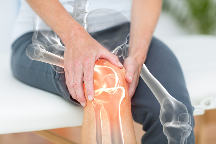
Knee pain has a way of interrupting everyday life. Whether it strikes during your morning walk, climbing the stairs, standing for long periods or even while trying to stay active, most people immediately assume arthritis is to blame. 

While joint damage is certainly a common cause of knee discomfort, it is far from the only explanation. In some cases, persistent knee pain may actually be linked to poor circulation or an underlying vascular condition that limits healthy blood flow to your legs. Understanding the difference is important because treating the wrong problem can delay relief and allow a more serious condition to progress. 

If you’ve been searching for answers about knee pain that isn’t arthritis, leg pain caused by poor circulation or wondering whether your symptoms could be related to vascular disease, learning the warning signs can help you take the next step toward an accurate diagnosis and effective treatment.

Why Knee Pain Isn’t Always a Joint Problem

When knee pain lingers despite rest, physical therapy or arthritis treatments, it may be time to look beyond the joint itself. Your knees rely on healthy blood flow to deliver oxygen and nutrients to the surrounding muscles, ligaments, tendons and other tissues. When circulation is reduced, or blood vessels become narrowed, blocked or inflamed, pain can develop even if the knee joint appears relatively healthy.

Because vascular conditions and orthopedic problems often share similar symptoms, many people spend months treating the wrong condition before discovering the true cause. Knowing when circulation may be involved can help you get the right diagnosis sooner.

Signs Your Knee Pain Could Be Vascular

While vascular-related knee pain doesn’t always feel the same as arthritis, there are several symptoms that may suggest poor circulation or another vascular condition is contributing to your discomfort.

Watch for:

  • Pain that worsens while walking and improves with rest
  • Cramping, aching or tightness in the calf, thigh or knee during activity
  • Cold feet or lower legs
  • Numbness or tingling in the legs
  • Slow-healing cuts or sores on the feet or lower legs
  • Skin discoloration or shiny skin on the legs
  • Weak or absent pulses in the feet
  • One leg that feels noticeably cooler than the other

These symptoms may indicate reduced blood flow caused by peripheral artery disease (PAD) or another vascular condition rather than a problem within the knee joint itself.

Common Vascular Conditions That May Cause Knee or Leg Pain

Several vascular disorders can create symptoms that mimic orthopedic injuries or arthritis.

Peripheral Artery Disease (PAD)

Peripheral artery disease occurs when plaque builds up inside the arteries, reducing blood flow to the legs. One of the earliest signs is claudication, which causes aching, cramping or fatigue in the legs during walking or exercise that improves with rest.

Although many people notice pain in the calf, discomfort can also occur around the knee, thigh or buttocks, depending on which arteries are affected.

Chronic Venous Insufficiency

Veins are responsible for returning blood to the heart. When vein valves weaken, blood can pool in the legs, leading to:

  • Aching or heaviness
  • Swelling around the lower legs
  • Leg fatigue
  • Pressure or discomfort that may extend toward the knees

Symptoms often worsen after prolonged standing and improve with elevation.

Popliteal Artery Problems

The popliteal artery runs directly behind the knee. Narrowing, compression or aneurysms involving this artery can sometimes cause pain, cramping or reduced circulation around the knee and lower leg, particularly during physical activity.

When Should You See a Vascular Specialist?

If knee pain persists despite traditional orthopedic care, or if you notice symptoms of poor circulation, don’t ignore them. Early diagnosis can help prevent complications while improving your treatment options.

Consider scheduling a vascular evaluation if you experience:

  • Pain while walking that consistently improves with rest
  • Leg cramps during activity
  • Persistent cold feet
  • Leg wounds that heal slowly
  • Skin color changes
  • Weak pulses in the feet
  • Ongoing knee or leg pain without a clear orthopedic diagnosis

A comprehensive vascular evaluation may include a physical examination, ankle-brachial index (ABI) testing or noninvasive vascular ultrasound to determine whether blood flow is contributing to your symptoms.

Find Answers at The Vascular Care Group

Knee pain isn’t always caused by arthritis, and understanding the difference can make all the difference in finding lasting relief. At The Vascular Care Group, our experienced vascular specialists evaluate a wide range of circulation-related conditions affecting the legs and work closely with patients to identify the source of their symptoms. 

If you’ve been living with persistent knee pain, leg discomfort or symptoms of poor circulation, schedule a consultation at one of our convenient locations to receive a personalized evaluation and explore the treatment options that may be right for you.

AAA (Abdominal Aortic Aneurysm): The Condition Most Patients Don’t Know They Have

Most people have never heard of an abdominal aortic aneurysm (AAA) until someone they know is diagnosed with one, yet this potentially life-threatening condition affects thousands of Americans every year and often develops without causing any noticeable symptoms. Because an abdominal aortic aneurysm can grow silently over time, many people don’t realize they have one until it is discovered during an imaging test for an unrelated health concern or, in more serious cases, after it ruptures and becomes a medical emergency. 

Understanding what an AAA is, who is most at risk and when to seek vascular screening can make a critical difference in preventing serious complications. In this guide, we’ll explain the causes, symptoms, risk factors, diagnosis and treatment options for abdominal aortic aneurysms, while highlighting why early detection and expert vascular care are essential for protecting your long-term health.

 Whether you have a family history of aneurysms, smoke or smoked in the past, or simply want to better understand your vascular health, knowing the facts about AAA could help save your life.

What Is an Abdominal Aortic Aneurysm (AAA)?

An abdominal aortic aneurysm (AAA) occurs when a section of the abdominal aorta, the body’s largest artery, becomes weakened and begins to bulge outward. The aorta carries oxygen-rich blood from the heart to the rest of the body, making it one of the most important blood vessels in your circulatory system.

As an aneurysm grows, the arterial wall becomes thinner and more fragile. If it ruptures, it can cause severe internal bleeding that requires immediate emergency medical care. The good news is that many abdominal aortic aneurysms can be detected early through routine screening and managed before they become life-threatening.

Who Is Most at Risk for an AAA?

Although anyone can develop an abdominal aortic aneurysm, certain factors significantly increase the risk.

Common risk factors include:

  • Adults over the age of 65
  • Current or former smokers
  • High blood pressure (hypertension)
  • High cholesterol
  • A family history of abdominal aortic aneurysms
  • Atherosclerosis (hardening of the arteries)
  • Being male, although women can also develop AAAs

If you fall into one or more of these categories, speaking with a vascular specialist about screening may be an important step toward protecting your health.

Why AAAs Are Often Called the “Silent Threat”

One of the most dangerous aspects of an abdominal aortic aneurysm is that it rarely causes symptoms during its early stages. Many patients feel perfectly healthy while the aneurysm gradually enlarges.

When symptoms do occur, they may include:

  • Persistent or deep abdominal pain
  • Back or flank pain
  • A noticeable pulsing sensation in the abdomen
  • Pain that does not improve with rest

A ruptured AAA is a medical emergency. Symptoms may include:

  • Sudden, severe abdominal or back pain
  • Dizziness or fainting
  • Rapid heartbeat
  • Low blood pressure
  • Loss of consciousness

If these symptoms develop, seek emergency medical attention immediately.

How Is an Abdominal Aortic Aneurysm Diagnosed?

Because AAAs often develop without symptoms, imaging plays a critical role in early detection.

Your vascular provider may recommend:

  • Abdominal ultrasound, the most common screening test
  • CT angiography (CTA) for detailed imaging
  • MRI in select cases
  • Imaging performed for unrelated medical conditions that unexpectedly reveals an aneurysm

Screening is especially valuable for individuals with multiple risk factors because it allows physicians to identify an aneurysm before it causes serious complications.

Treatment Depends on the Size of the Aneurysm

Not every abdominal aortic aneurysm requires immediate surgery. Your treatment plan depends on several factors, including the aneurysm’s size, growth rate and your overall health.

Treatment options may include:

Careful Monitoring

Small aneurysms may only require routine imaging and follow-up appointments to ensure they do not grow too quickly.

Lifestyle Changes

Your physician may recommend:

  • Quitting smoking
  • Managing blood pressure
  • Lowering cholesterol
  • Exercising safely
  • Following a heart-healthy diet

These steps support overall vascular health and may help reduce future complications.

Minimally Invasive Endovascular Repair (EVAR)

Many patients qualify for endovascular aneurysm repair (EVAR), a minimally invasive procedure that places a stent graft inside the weakened portion of the aorta. This approach often results in:

  • Smaller incisions
  • Less discomfort
  • Shorter hospital stays
  • Faster recovery compared to traditional surgery

Open Surgical Repair

For larger or more complex aneurysms, open surgery may be the safest and most effective treatment option.

Can an Abdominal Aortic Aneurysm Be Prevented?

While not every AAA can be prevented, maintaining good vascular health can significantly reduce your risk.

Healthy habits include:

  • Avoiding tobacco products
  • Keeping blood pressure under control
  • Managing diabetes
  • Maintaining healthy cholesterol levels
  • Staying physically active
  • Eating a balanced diet rich in fruits, vegetables and lean proteins
  • Keeping regular appointments with your primary care physician

For many high-risk adults, routine vascular screening offers the greatest opportunity to detect an aneurysm before it becomes dangerous.

When Should You See a Vascular Specialist?

Many patients don’t realize they should see a vascular specialist until symptoms appear, but waiting isn’t always the safest choice.

You should consider scheduling a vascular evaluation if you:

  • Have a family history of AAA
  • Are over age 65 with a history of smoking
  • Have been diagnosed with peripheral artery disease or other vascular conditions
  • Have been told an aneurysm was found during another imaging study
  • Have ongoing abdominal or back pain that cannot be explained

Early evaluation provides peace of mind and allows your care team to determine whether monitoring or treatment is appropriate.

Protect Your Vascular Health with The Vascular Care Group

Early detection remains one of the most effective ways to prevent complications from an abdominal aortic aneurysm. At The Vascular Care Group, our experienced vascular specialists provide comprehensive evaluations, advanced diagnostic imaging and personalized treatment plans for patients throughout the communities we serve. Whether you’re concerned about your risk factors, have been referred for vascular screening or need expert treatment for an abdominal aortic aneurysm, our team is here to help you take proactive steps toward better vascular health. 

Contact The Vascular Care Group today to schedule a consultation and learn whether an AAA screening is right for you.


Why Your Legs Feel Worse in Summer Heat (And What Your Circulation Is Telling You)

Have you noticed that your legs feel heavier, more swollen, or simply more uncomfortable when the temperatures rise? You are not imagining it. Summer heat can have a noticeable impact on your circulation, especially for people who already experience vein-related symptoms such as leg swelling, aching, cramping, fatigue, or visible varicose veins. 

As the body works to cool itself in warmer weather, blood vessels naturally expand, which can make it harder for blood to return efficiently from the legs back to the heart. For some individuals, this can lead to increased pressure in the veins and more noticeable discomfort throughout the day. 

Understanding why your legs feel worse in the summer can help you identify changes in your vascular health, recognize when symptoms may need attention, and take proactive steps to support better circulation. 

Why Summer Heat Can Make Your Legs Feel Worse

For many people, summer means spending more time outdoors, traveling, exercising, and enjoying warmer weather. However, higher temperatures can also bring unwanted changes, especially when it comes to your legs. If you notice that your legs feel more swollen, tired, achy, or heavy during the summer months, your circulation may be playing a role.

Heat affects the way your blood vessels function, and for people with underlying vein concerns, symptoms can become more noticeable. Whether you experience occasional discomfort or ongoing issues with swelling or varicose veins, understanding the connection between summer weather and vascular health can help you take the right steps toward relief.

How Does Hot Weather Affect Your Circulation?

When temperatures rise, your body naturally tries to cool itself by widening blood vessels near the surface of your skin. This process, known as vasodilation, helps release heat but can also affect how efficiently blood moves through your veins.

The veins in your legs already work against gravity to move blood back toward your heart. When veins become more relaxed in hot weather, blood may collect more easily in the lower legs, increasing pressure and contributing to discomfort.

Common summer-related circulation symptoms may include:

  • Swelling around the ankles, feet, or lower legs
  • A feeling of heaviness or tiredness in the legs
  • Aching or throbbing discomfort after standing or walking
  • More noticeable varicose veins
  • Tightness or changes in how your shoes or clothing fit

Why Leg Swelling Often Gets Worse in the Summer

Leg swelling, also known as edema, occurs when excess fluid builds up in the tissues. While occasional swelling can happen after a long day on your feet, frequent or worsening swelling may be a sign that your veins are having difficulty circulating blood properly.

Summer activities can sometimes make symptoms more noticeable. Long car rides, flights, extended periods of standing, and increased outdoor activity can all place additional stress on your circulation.

To help support healthy blood flow during warmer months, consider these simple habits:

  • Stay hydrated throughout the day
  • Avoid sitting or standing in one position for long periods
  • Take short walks to encourage circulation
  • Elevate your legs when resting
  • Wear comfortable clothing that does not restrict blood flow
  • Follow your healthcare provider’s recommendations for compression therapy if needed

Are Summer Leg Symptoms a Sign of a Vein Problem?

While heat can temporarily worsen circulation symptoms, it can also highlight underlying vascular conditions. Issues such as chronic venous insufficiency occur when the valves inside the veins do not work properly, allowing blood to flow backward and pool in the legs.

People with vein conditions may notice symptoms such as:

  • Persistent leg swelling
  • Aching or discomfort that improves with elevation
  • Skin changes around the ankles
  • Leg cramps or a feeling of pressure
  • Enlarged, twisted varicose veins

If your symptoms continue beyond the summer months or interfere with your daily activities, it may be worth speaking with a vascular specialist. Early evaluation can help identify the cause of your symptoms and determine appropriate treatment options.

When Should You See a Vascular Specialist?

Not every case of leg discomfort requires medical treatment, but certain warning signs should not be ignored. Seeking professional guidance can help protect your long-term vascular health and prevent symptoms from progressing.

Consider scheduling an evaluation if you experience:

  • Swelling that occurs regularly or affects one leg more than the other
  • Leg pain, heaviness, or fatigue that impacts your routine
  • Skin discoloration or changes near the ankles
  • Non-healing sores or wounds on your legs
  • Symptoms that continue despite lifestyle changes

A vascular specialist can assess your circulation, identify potential vein issues, and recommend personalized treatment options designed around your needs.

Find Expert Vascular Care at The Vascular Care Group

If summer heat is making your legs feel heavier, more swollen, or more uncomfortable, The Vascular Care Group is here to help. Serving communities throughout Connecticut, Maine, and Massachusetts, our experienced vascular specialists provide comprehensive care for a wide range of vein and vascular conditions. From evaluating circulation concerns to offering advanced treatment options, our team focuses on helping patients improve their vascular health and quality of life. 

Contact The Vascular Care Group today to schedule a consultation and take the first step toward healthier, more comfortable legs.


Muscle Pain or Vein Pain? How to Tell the Difference

Most people have experienced leg pain at some point: whether after a long day on their feet, an intense workout, or simply waking up with an unexplained ache. In many cases, the discomfort is caused by a strained muscle or minor injury that improves with time and rest. But what happens when the pain lingers, returns regularly, or is accompanied by symptoms like swelling, heaviness, throbbing, or visible veins? 

While muscle pain and vein pain can sometimes feel similar, they often have very different underlying causes. Understanding the difference between muscle pain and vascular pain is important because certain vein conditions can signal circulation problems that may worsen without proper treatment. 

Knowing what signs to watch for can help you determine whether your symptoms are related to a muscle injury or a vascular condition and when it may be time to seek expert medical evaluation. 

What Does Muscle Pain Typically Feel Like?

Muscle pain is one of the most common causes of discomfort in the legs. It often develops after physical activity, overuse, repetitive movements, or minor injuries. In most cases, muscle pain improves gradually as the body heals.

Common signs of muscle-related pain include:

  • Soreness after exercise or physical activity
  • Tenderness when touching the affected area
  • Cramping or tightness in the muscle
  • Pain that improves with rest
  • Discomfort that is limited to a specific muscle group

Muscle strains and overuse injuries are usually temporary and tend to follow a predictable healing process. While the pain may be uncomfortable, it generally improves over days or weeks rather than progressively worsening.

How Vein Pain Is Different

Vein pain is often caused by problems within the circulatory system rather than the muscles themselves. When veins have difficulty returning blood to the heart, pressure can build up in the legs, leading to discomfort and other symptoms.

Unlike muscle pain, vascular pain is often described as:

  • Aching or throbbing
  • A feeling of heaviness in the legs
  • Burning or itching sensations
  • Swelling around the ankles or calves
  • Symptoms that worsen after prolonged standing or sitting

Many people with vein disease notice that their legs feel worse at the end of the day and better after elevating their legs. This pattern can be an important clue that the issue may be related to circulation rather than a muscle injury.

Warning Signs That May Point to a Vascular Condition

While not every case of leg pain is serious, certain symptoms should not be ignored. Vein-related conditions can progress over time and may increase the risk of complications if left untreated.

You may want to schedule an evaluation with a vascular specialist if you experience:

  • Persistent leg pain that does not improve with rest
  • Leg heaviness or fatigue during normal daily activities
  • Visible varicose veins or spider veins
  • Swelling that regularly affects one or both legs
  • Skin discoloration around the ankles
  • Non-healing wounds or ulcers on the lower legs
  • Pain that worsens after standing for long periods

These symptoms may be associated with chronic venous insufficiency, varicose veins, or other vascular conditions that affect healthy blood flow.

When Leg Pain Could Be an Emergency

Although many vascular issues develop gradually, some symptoms require immediate medical attention.

Seek emergency care if you experience:

  • Sudden swelling in one leg
  • Severe pain that develops quickly
  • Redness and warmth in the affected area
  • Shortness of breath or chest pain accompanied by leg symptoms

These signs may indicate a blood clot or another serious vascular condition that requires prompt treatment.

Why an Accurate Diagnosis Matters

Because muscle pain and vein pain can sometimes feel similar, self-diagnosing can be difficult. A person may assume they are dealing with a lingering muscle strain when the real issue is related to circulation.

An accurate diagnosis helps ensure that you receive the appropriate treatment and avoid unnecessary discomfort. Vascular specialists use advanced diagnostic tools, including ultrasound imaging, to evaluate blood flow and identify the source of leg pain. The sooner a vascular condition is detected, the more treatment options may be available.

Don’t Ignore Persistent Leg Pain

Leg discomfort is often easy to dismiss, especially when life gets busy. However, pain, swelling, heaviness, or visible vein changes can sometimes be signs of an underlying vascular condition that deserves medical attention. Understanding the difference between muscle pain and vein pain can help you take the next step toward protecting your long-term vascular health.

Get Expert Vascular Care in New England 

If you’re experiencing ongoing leg pain, swelling, heaviness, or other symptoms that may be related to circulation, the experts at TVCG are here to help. Our team specializes in diagnosing and treating complex vascular conditions for patients across New England. Using advanced diagnostic technology and personalized treatment plans, we help patients find answers, improve circulation, and regain confidence in their health. 

Contact  TVCG today to schedule a consultation with a vascular specialist.


From Workdays to Weekends: How Men’s Daily Habits Impact Circulation

For many men, the long work week to an active weekend can be a challenge. Whether your Monday through Friday involves sitting behind a desk or physical labor on a job site, your habits are quietly shaping your vascular health. At The Vascular Care Group, world-class vascular care begins with understanding how these routines affect your circulation.

Maintaining healthy blood flow is essential for peak performance, both professionally and personally. Here is how your daily choices impact your veins and arteries, and how our comprehensive services can help you stay ahead of vascular disease.

The Sedentary Workday: A Circulation Killer

In today’s professional world, many men spend almost eight hours a day seated. This lengthy sitting causes blood to pool in the lower extremities, increasing pressure on the vein walls and valves. Over time, this can lead to Chronic Venous Insufficiency (CVI) or even the development of Deep Vein Thrombosis (DVT).

If your workday is stationary, your “muscle pump”, the calf muscles that help push blood back to the heart, remains inactive. We recommend integrating small movements into your routine, such as brief walks or calf raises, to stimulate flow and reduce the risk of long-term damage.

Overexertion and Arterial Strain

When the weekend arrives, some men shift into active mode, like engaging in heavy outdoor work or more intense workouts. While physical activity is crucial for cardiovascular health, sudden, intense exertion after an inactive week can strain the arterial system, especially if underlying plaque or narrowing is present.

Proper circulation ensures that oxygen reaches your muscles efficiently. If you experience persistent cramping, heaviness, or “claudication” (pain during exercise that stops with rest), it may be a sign of Peripheral Artery Disease (PAD). Recognizing these signals early is the key to getting back to doing the things you love without limitations.

Habits Beyond the Clock: Diet, Hydration, and Recovery

Circulation isn’t just about movement; it’s about the “fuel” you provide your vascular system.

  • Sodium and Hydration: High-sodium “on-the-go” lunches can lead to fluid retention and increased blood pressure, straining the delicate lining of your vessels.
  • Nicotine Use: Smoking and vaping are among the most significant risk factors for arterial hardening and blockages.
  • Alcohol Consumption: Excessive weekend drinking can lead to dehydration and erratic heart rhythms, both of which complicate healthy circulation.

Comprehensive Care in New England

At The Vascular Care Group, we provide comfortable, convenient, and authoritative medical guidance to men throughout the New England area. Our advanced technologies, including non-invasive vascular ultrasound and vascular testing, allow us to assess your circulatory health with precision.

Don’t let poor circulation sideline your goals. Contact The Vascular Care Group today to schedule a consultation and ensure your vascular health supports your lifestyle.